Merkliste
Die Merkliste ist leer.
Der Warenkorb ist leer.
Kostenloser Versand möglich
Bitte warten - die Druckansicht der Seite wird vorbereitet.
Der Druckdialog öffnet sich, sobald die Seite vollständig geladen wurde.
Sollte die Druckvorschau unvollständig sein, bitte schliessen und "Erneut drucken" wählen.

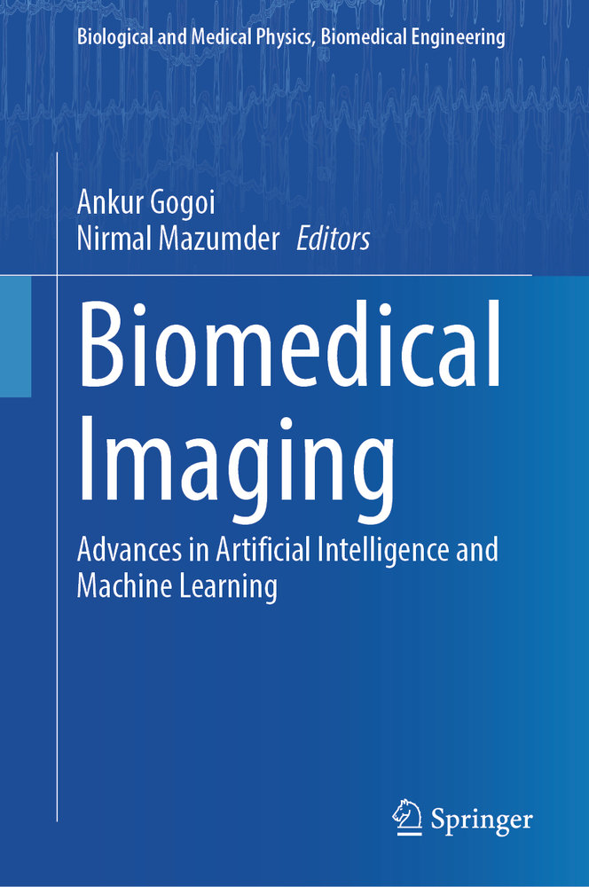
Biomedical Imaging

Advances in Artificial Intelligence and Machine Learning
BuchGebunden
EUR160,00

Produktbeschreibung

This book presents the rapidly developing field of artificial intelligence and machine learning and its application in biomedical imaging. As is known, starting from the diagnosis of fractures by using X-rays to understanding the complex structure and function of the brain, biomedical imaging has contributed immensely toward the development of precision diagnosis and treatment strategies for numerous diseases. While continuous evolution in imaging technologies have enabled the acquisition of images having resolution and contrast far better than ever, it significantly increased the volume of data associated with each image scan-making it increasingly difficult for experts to analyze and interpret. In this context, the application of artificial intelligence (AI) and machine learning (ML) tools has become one of the most exciting frontlines of contemporary research in biomedical imaging due to their capability to extract minute traces of various disease signatures from large and complicated datasets and providing clear insight into the potential abnormalities with excellent accuracy, sensitivity, and specificity. The hallmark of this book will be the contributions from international leaders on different AI-aided advanced biomedical imaging modalities and techniques. Included will be comprehensive description of several of the technology-driven spectacular advances made over the past few years that have allowed early detection and delineation of abnormalities with sub-pixel image segmentation and classification. Starting from the fundamentals of biomedical image processing, the book presents a streamlined and focused coverage of the core principles, theoretical and experimental approaches, and state-of-the-art applications of most of the currently used biomedical imaging techniques powered by AI.
Weiterlesen

Details

ISBN/GTIN978-981-97-5344-4
ProduktartBuch
EinbandGebunden
VerlagSpringer
ErscheinungsortSingapore
ErscheinungslandSingapur
Erscheinungsdatum27.11.2024
Auflage2024
Seiten330 Seiten
SpracheEnglisch
Illustrationen16 s/w Abbildungen, 88 farbige Abbildungen
Artikel-Nr.29039689
KatalogVLB
Datenquelle-Nr.478331a66eda4531bc87c132601ea122
Weitere Details

Reihe

Bewertungen

Autor/in


Dr. Ankur Gogoi is an assistant professor in the Department of Physics at Jagannath Barooah University (formerly Jagannath Barooah College), Assam, India. He completed his Ph.D. in 2012 from Tezpur University, Assam, India, and worked as a postdoctoral research associate at the Institute of Biophotonics, National Yang Ming Chiao Tung University, Taiwan from 2016 to 2018. Dr. Gogoi has also held the position of assistant professor of Physics at The Assam Kaziranga University, Jorhat, India and Girijananda Chowdhury Institute of Management and Technology-Tezpur, Assam, India. His research interests are in optoelectronic instrumentation, linear and nonlinear optical microscopy, photonics, and nanotechnology.


Dr. Nirmal Mazumder is an associate professor at the Department of Biophysics, Manipal School of Life Sciences, Manipal Academy of Higher Education (MAHE), Manipal, India. He obtained his Ph.D. in 2013 from National Yang Ming Chiao Tung University, Taipei, Taiwan. From 2013 to 2016, he worked as a postdoctoral fellow at the University of Virginia, the USA, and the Italian Institute of Technology, Genoa, Italy. He has been developing nonlinear optical microscopes, including two photon fluorescence, second harmonic generation, and coherent anti-Stokes Raman scattering for biomedical applications. He works extensively on the biophysical aspects of starch, including various spectroscopy and microscopy techniques.

Schlagworte